How to monitor the ECG
Choose all the options that are correct and then select Confirm.

That is not quite right.
No, that is still not quite right. All the statements were true.
Some rhythms may be visible clearly in only one or two of the leads on a 12-lead ECG. Self-adhesive pads can usually be applied more quickly than other electrodes and permit prompt defibrillation if cardiac arrest with a shockable rhythm occurs, but monitoring should not be delayed if self-adhesive pads and a defibrillator are not available immediately and other forms of monitoring are. If ECG electrodes are placed over muscle there will be more likelihood of a poor quality ECG due to muscle artefact. If monitoring is needed it should be established as soon as possible with whatever equipment is available. To achieve adequate quality ECG signals it may be necessary to remove excess body hair, as well as sweat or grease from the skin, before applying the electrodes.
That is not right.
Your answers are still not correct. All the statements were true.
Some rhythms may be visible clearly in only one or two of the leads on a 12-lead ECG. Self-adhesive pads can usually be applied more quickly than other electrodes and permit prompt defibrillation if cardiac arrest with a shockable rhythm occurs, but monitoring should not be delayed if self-adhesive pads and a defibrillator are not available immediately and other forms of monitoring are. If ECG electrodes are placed over muscle there will be more likelihood of a poor quality ECG due to muscle artefact. If monitoring is needed it should be established as soon as possible with whatever equipment is available. To achieve adequate quality ECG signals it may be necessary to remove excess body hair, as well as sweat or grease from the skin, before applying the electrodes.
Yes, that is right. Some rhythms may be visible clearly in only one or two of the leads on a 12-lead ECG. Self-adhesive pads can usually be applied more quickly than other electrodes and permit prompt defibrillation if cardiac arrest with a shockable rhythm occurs, but monitoring should not be delayed if self-adhesive pads and a defibrillator are not available immediately and other forms of monitoring are. If ECG electrodes are placed over muscle there will be more likelihood of a poor quality ECG due to muscle artefact. If monitoring is needed it should be established as soon as possible with whatever equipment is available. To achieve adequate quality ECG signals it may be necessary to remove excess body hair, as well as sweat or grease from the skin, before applying the electrodes.
References
See chapter 8 of the ALS manual for further explanation and examples about how to analyse ECG rhythm strips.
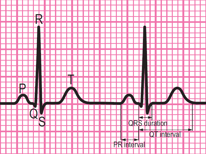
Components of a normal ECG complex
- Depolarisation begins in the SA node and then spreads through to the atrial myocardium
- The heart reacts to this depolarisation. The atrial contraction is recorded on the rhythm strip as the P wave
- The small isoelectric segment between the P wave and QRS complex represents the delay in transmission through the AV node
- Depolarisation of the bundle of His, bundle branches and ventricular myocardium is shown on the rhythm strip as the QRS complex
- The T wave represents recovery of the resting potential in the cells of the conducting system and ventricular myocardium


The 6-stage approach
1. Is there any electrical activity?
2. What is the ventricular (QRS) rate?
3. Is the QRS rhythm regular or irregular?
4. Is the QRS width normal (narrow) or broad?
Any cardiac rhythm can be described accurately and managed safely and effectively using the first four steps.
5. Is atrial activity present? (If so, what is it: Typical sinus P waves? Atrial fibrillation? Atrial flutter? Abnormal P waves?)
6. How is atrial activity related to ventricular activity? (e.g 1:1 conduction, 2:1 conduction, etc, or no relationship)


