Acute ST-segment-elevation anterolateral myocardial infarction
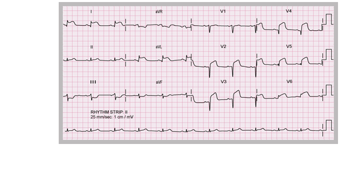

There is ST segment elevation in the anterior chest leads and in the lateral leads, so this is an anterolateral ST elevation MI and immediate reperfusion therapy should be considered.
Look at this ECG from a patient with severe chest pain. Click on the coloured arrows to find out more. Select Next to continue.
Note also that there are Q waves in the anterior leads. When an area of myocardium is damaged to the point where it becomes electrically inactive the overlying ECG leads show the development of Q waves. These can appear very early in the course of infarction, but may develop later due to progressive damage. Early intervention to re-open a blocked coronary artery may limit the amount of myocardial damage and reduce or prevent the development of Q waves.
Look at this ECG from a patient with severe chest pain. Click on the coloured arrows to find out more. Select Next to continue.
Look at this ECG from a patient with severe chest pain. Click on the coloured arrows to find out more. Select Next to continue.

Look at this ECG from a patient with severe chest pain. Click on the coloured arrows to find out more. Select Next to continue.


References

Acute coronary syndromes: immediate treatment and
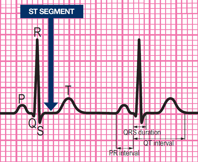
components of a normal ECG rhythm strip
Acute coronary syndromes: immediate treatment
Give immediate treatment to relieve symptoms, limits myocardial damage and reduce the risk of cardiac arrest. Immediate general treatment for ACS comprises:
- Aspirin
- Nitroglycerine
- Oxygen
- Morphine or Diamorphine
Components of a normal ECG complex
- Depolarisation begins in the SA node and then spreads through the atrial myocardium
- This depolarisation is recorded on the rhythm strip as the P wave. The heart responds to this electrical stimulus byatrial contraction
- The small isoelectric segment between the P wave and QRS complex represents the delay in transmission through the AV node
- Depolarisation of the bundle of His, bundle branches and ventricular myocardium is shown on the rhythm strip as the QRS complex
- The T wave represents recovery of the resting potential (repolarisation) in the cells of the conducting system and ventricular myocardium


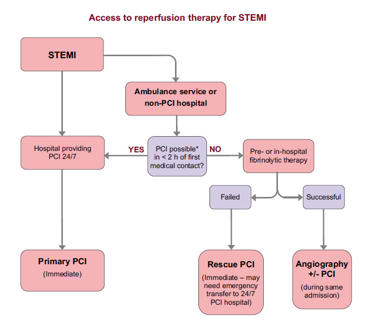
The ABCDE approach and access to reperfusion therapy for STEMI
All patients with a suspected acute coronary syndrome should be assessed using the ABCDE approach.
The ABCDE approach stands for:
- Airway
- Breathing
- Circulation
- Disability
- Exposure
In addition where an ST-elevation MI is evident immediate reperfusion therapy should be arranged unless contra-indicated.