
12-lead ECG in acute myocardial infarction
The appearance of a 12-lead ECG can help to identify the presence of acute myocardial infarction and to determine the appropriate treatment.
When a patient presents with a history suggestive of an acute coronary syndrome a 12-lead ECG should be recorded as soon as possible. The features seen on the ECG will help to confirm the diagnosis in many cases and in all cases will dictate the immediate choice of treatment that should be used.
The following examples will illustrate some of the typical patterns of ECG abnormality that are seen in acute MI.
Select Next to continue.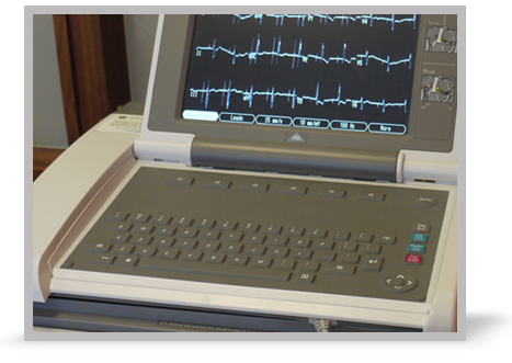


References

See chapter 4 of the ALS manual for further detail relating to the assessment and treatment of Acute Coronary Syndromes
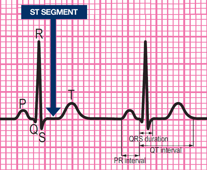
Components of a normal ECG complex
- Depolarisation begins in the SA node and then spreads through the atrial myocardium
- This depolarisation is recorded on the rhythm strip as the P wave. The heart responds to this electrical stimulus byatrial contraction
- The small isoelectric segment between the P wave and QRS complex represents the delay in transmission through the AV node
- Depolarisation of the bundle of His, bundle branches and ventricular myocardium is shown on the rhythm strip as the QRS complex
- The T wave represents recovery of the resting potential (repolarisation) in the cells of the conducting system and ventricular myocardium


Settings
Font colour
default inverted high contrast high contrast inverted high contrast soft green on blackSample text
text looks like thisTEXT LOOKS LIKE THIS