How do I understand the basic physiology of the ECG?
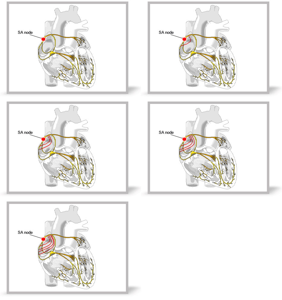
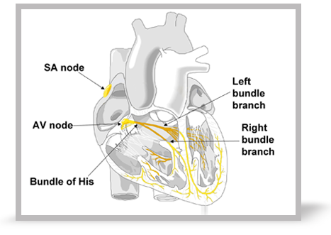
In normal sinus rhythm, depolarisation begins in a group of specialist pacemaker cells called the sino-atrial (SA) node.
A wave of depolarisation then spreads from the SA node through the atrial myocardium. This is seen on the ECG as the P wave.
Atrial contraction is the mechanical response to this electrical impulse.
Select More to continue.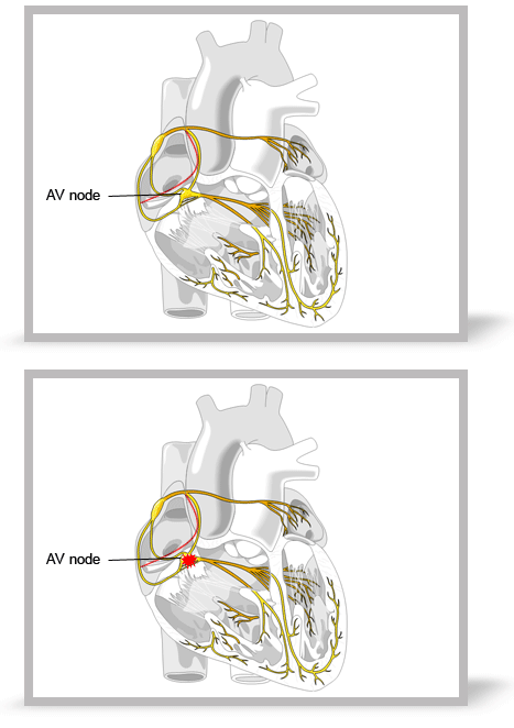
When the electrical impulse in the atria reaches the AV node it is conducted slowly, represented on the ECG largely by the isoelectric portion of the PR interval. When the impulse leaves the AV node there is rapid conduction to the ventricular myocardium by specialist conducting tissue (Purkinje fibres).
Select More to continue.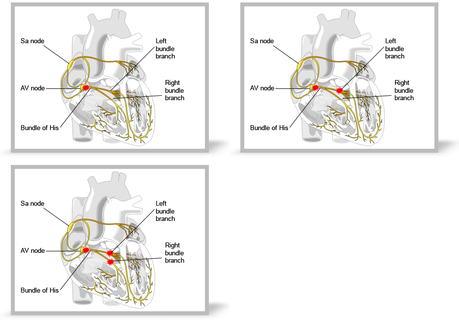
The bundle of His carries the Purkinje fibres from the AV node and then divides into right and left bundle branches, spreading out through the right and left ventricles respectively.
Rapid conduction down these fibres ensures that the ventricles contract in a co-ordinated fashion. Depolarisation of the bundle of His, bundle branches and ventricular myocardium is seen on the ECG as the QRS complex.
Ventricular contraction is the mechanical response to this electrical impulse.
Select Restart to play the animation again or select Next to continue.Components of a normal ECG complex
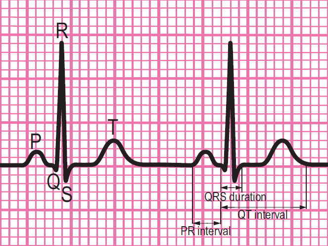
- Depolarisation begins in the SA node and then spreads through the atrial myocardium
- This depolarisation is recorded on the rhythm strip as the P wave. The heart responds to this electrical stimulus byatrial contraction
- The small isoelectric segment between the P wave and QRS complex represents the delay in transmission through the AV node
- Depolarisation of the bundle of His, bundle branches and ventricular myocardium is shown on the rhythm strip as the QRS complex
- The T wave represents recovery of the resting potential (repolarisation) in the cells of the conducting system and ventricular myocardium


The 6-stage approach
1. Is there any electrical activity?
2. What is the ventricular (QRS) rate?
3. Is the QRS rhythm regular or irregular?
4. Is the QRS width normal (narrow) or broad?
Any cardiac rhythm can be described accurately and managed safely and effectively using the first four steps.
5. Is atrial activity present? (If so, what is it: Typical sinus P waves? Atrial fibrillation? Atrial flutter? Abnormal P waves?)
6. How is atrial activity related to ventricular activity? (e.g 1:1 conduction, 2:1 conduction, etc, or no relationship)

