
How do I monitor the ECG?
Where a decision has been taken to monitor the ECG there are three options as to the method of monitoring to be used.
Select each number to find out more.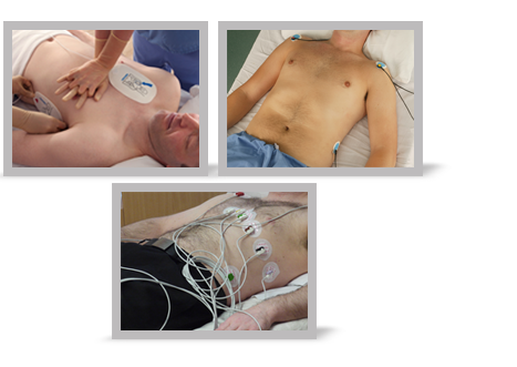

Self-adhesive pads are used in emergencies, such as when a patient has collapsed. The cardiac rhythm can be assessed as soon as possible by applying defibrillator pads to the patient.
The self-adhesive pads can be used for monitoring rhythm and for hands-free shock delivery. They should be applied beneath the right clavicle and in the left mid-axillary line, overlying the V6 ECG electrode position. Alternatively anterior and posterior positions can be used.
Select each number to find out more. Select Menu to return to the main menu.

If a patient requires monitoring, but is not so critically ill that defibrillator pads are likely to be needed, 3-lead monitoring can be used to observe their cardiac rhythm. This is the standard form of ECG monitoring employed by many cardiac monitors and defibrillators in general clinical use. Note that the electrodes are placed over the bony parts of the shoulders and usually over the pelvic crest, to minimise artefact from underlying muscle.
Select each number to find out more. Select Menu to return to the main menu.

In some clinical settings, such as a cardiac care unit, 12-lead monitoring is available to enable early detection of ECG abnormalities that may develop in a limited number of leads. In particular this may help to identify some rhythm abnormalities. It is not always possible to identify an arrhythmia from a single lead ECG recording and occasionally the precise nature of the arrhythmia may be visible in only one of the 12 conventional ECG leads. In these cases 12-lead ECG monitoring may identify the cardiac rhythm accurately. Alternatively a 12-lead ECG can be recorded during an arrhythmia that has been detected by simpler forms of monitoring.
The heart is a three-dimensional organ and the 12-lead ECG addresses this by examining the heart’s electrical signals from 12 different directions.
Select each number to find out more. Select Menu to return to the main menu.


References


The 6-stage approach
1. Is there any electrical activity?
2. What is the ventricular (QRS) rate?
3. Is the QRS rhythm regular or irregular?
4. Is the QRS width normal (narrow) or broad?
Any cardiac rhythm can be described accurately and managed safely and effectively using the first four steps.
5. Is atrial activity present? (If so, what is it: Typical sinus P waves? Atrial fibrillation? Atrial flutter? Abnormal P waves?)
6. How is atrial activity related to ventricular activity? (e.g 1:1 conduction, 2:1 conduction, etc, or no relationship)